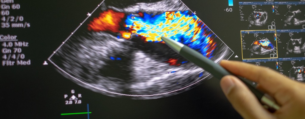
Echocardiogram Reads And Interpretations $45 Per Study
NDI rates for interpreting the results of echocardiogram (echo) tests of the heart and blood vessels start at $45 per study.
How You Request An Echocardiogram Scan Interpretation Or Echo Reading Services
Call 216-514-1199, email info@ndximaging.com or submit the form here.
Please contact NDI to request a diagnostic interpretation of an echocardiogram scan or echo reading services.
US Telecardiology Service To Diagnose Heart Disease
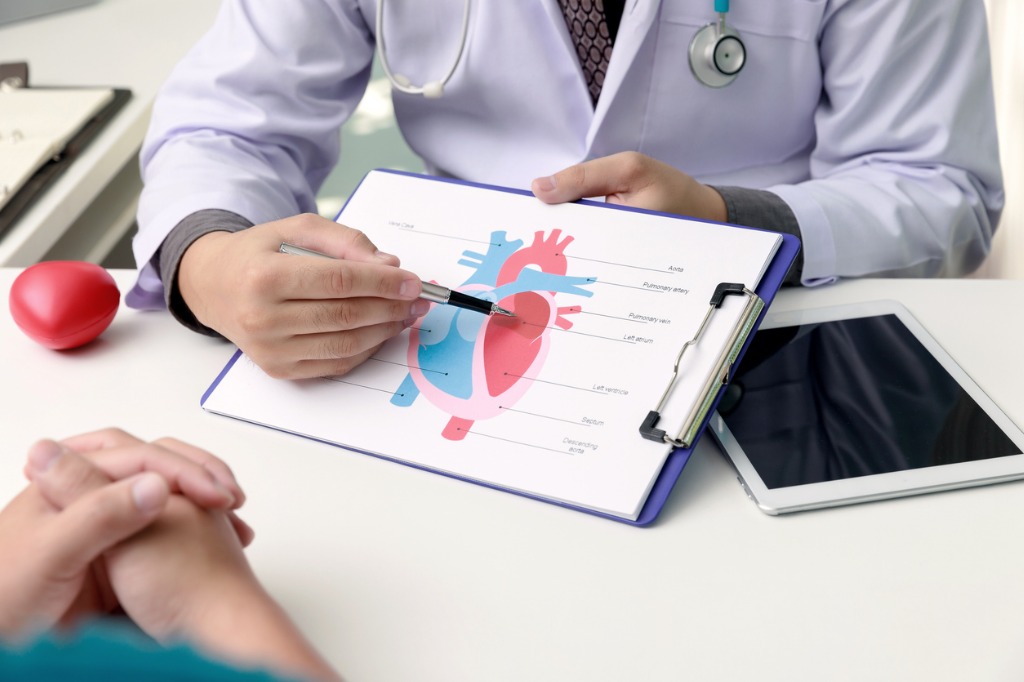
The NDI telecardiology company provides remote online reporting of cardiac imaging studies of the circulatory system, cardiovascular system, heart and blood vessels. NDI telecardiology services are used by U.S. cardiovascular imaging centers to receive remote interpretations of electrocardiogram (ECG or EKG) results from heart imaging specialists online.
About NDI Echocardiogram Reading Services
NDI is a teleradiology company that provides telecardiology services, radiology reads, remote cardiology reading services and echocardiogram (echocardiography) reading services for healthcare facilities in all 50 states in the USA.
The easiest way to get access to teleradiology services in the United States is to call National Diagnostic Imaging at 1-800-950-5257 or email info@ndximaging.com.
Radiologists at National Diagnostic Imaging submit their detailed and comprehensive echocardiogram findings and interpretations to referring physicians via a PACS and teleradiology.
NDI doctors are U.S. Board Certified and hold certifications in Echocardiography. We provide interpretations for cardiologists, cardiology clinics, hospitals, private physicians, urgent care centers, internal medicine departments, cardiac surgeons and heart specialists.
Healthcare facilities that contract with NDI for teleradiology services use echocardiograms to see a patient’s heart beating and pumping blood. NDI clients use the images from an echocardiogram to identify heart disease.
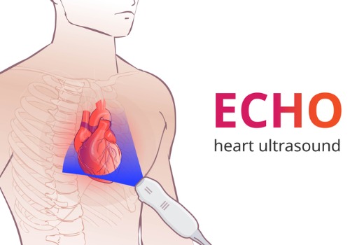
An echocardiography, echocardiogram, cardiac echo or simply an echo, is an ultrasound of the heart. Echo tests are used to monitor congenital heart disease and pulmonary hypertension.
An echocardiogram, unlike an ECG, provides a detailed image of the heart muscle and heart valves. Learn how to read an echocardiogram report, here.
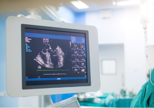
Echocardiography is one of the most widely used diagnostic imaging modalities in cardiology. An echocardiogram provides information about how blood flows through the different parts of the heart and major blood vessels.
The Intersocietal Accreditation Commission (IAC) sets standards for echo labs across the US.
NDI Provides Echo Reads And Final Reports To Referring Physicians
NDI fellowship trained and US board certified subspecialty trained radiologists read echocardiogram to help doctors find out if their patient has a heart murmur, atrial fibrillation or valve problems.

NDI interprets echocardiograms for cardiologist who are experts in the care of their patient’s heart and blood vessels.
Echo reads and reports submitted by NDI cardiologists via teleradiology through a PACS system help doctors treat or help prevent a number of cardiovascular problems.
NDI teleradiologists interpret echocardiogram scans while not physically present in the location where the images are generated.

Hospitals, other cardiologists mobile imaging companies, radiology departments, urgent care facilities and private practices use teleradiology services provided by National Diagnostic Imaging.
Cardiologists for National Diagnostic Imaging interpret and report on echocardiogram images via teleradiology to assist referring physicians that diagnose heart conditions.
NDI teleradiologists use echocardiogram scans to detect fluid around the heart, thickening of the heart tissue or clotting.
NDI subspecialty trained cardiologists know how to accurately interpret echocardiograms. Echocardiography is an important tool in assessing wall motion abnormality in patients with suspected cardiac disease. NDI customers use echocardiograms to generate images of the heart of a patient in real-time using ultrasound.
NDI Echocardiogram Reading Fees
Rates for echo reads, in volume, for healthcare facilities in the United States start at $28. Get more information on teleradiology reading service pricing for organizations, here.
Rates for remote echocardiography reading services start at $28 per study. NDI currently provides tele-echocardiography and remote cardiology reading services to radiology facilities all across the United States.
Cardiology image reading services and interpretations services start at $13 per study based on the imaging modality and volume.
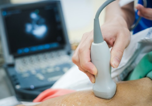
Echocardiography is a type of medical imaging of the heart, using standard ultrasound or Doppler ultrasound. An “echo” is a graphic outline of the heart’s movement.
During an echo test, NDI customers use ultrasound (high-frequency sound waves) from a hand-held wand placed on their patient’s chest to take pictures of the heart’s valves and chambers.
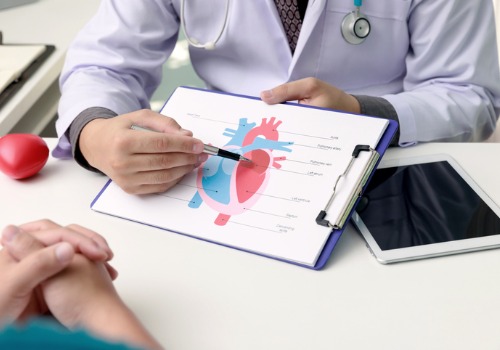
Types of Echocardiography
- Transthoracic echocardiogram
- Transesophageal echocardiogram
- Stress echocardiography
- Intracardiac echocardiography
- Intravascular ultrasound
NDI cardiologists interpret the following studies via teleradiology and submit their findings to referring physicians via a PACS, and in some cases, by email:
- Echocardiogram
- ECG
- Stress ECG
- Nuclear Stress Test (Including Stress ECG)
- Stress Echocardiogram (Including Stress ECG)
- Coronary CTA
- Cardiac MRI
- Fetal Echocardiogram
A fetal echocardiogram (also called a fetal echo) uses sound waves to create pictures of an unborn baby’s heart. This ultrasound test shows the structure of the heart and how well it’s working.
The fetal echocardiogram test is performed by a specially trained ultrasound sonographer and the images are interpreted by a pediatric cardiologist who specializes in fetal congenital heart disease.
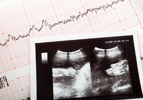
Interpreting An Echocardiogram Report
An echocardiogram report describes the heart’s structures in detail and often includes a summary of the cardiologist’s findings.
Information Included In An Echocardiogram Report
- The size of the heart chambers and thickness of the heart muscle
- The function of the left and right ventricles (pumping chambers)
- A description of the shape, movement, and function of the heart valves
- A description of other structures that are important for heart function, including the large arteries and veins, the lining of the heart (the pericardium), and any abnormalities, such as blood clots
Interpreting The Results Of An Echocardiogram
The resulting image of an echocardiogram can show a big picture image of heart health, function, and strength. For example, the test can show if the heart is enlarged or has thickened walls. Walls thicker than 1.5cm are considered abnormal. They may indicate high blood pressure and weak or damaged valves.
The echocardiogram can help detect:
- Abnormal heart valves
- Congenital heart disease (abnormalities present at birth)
- Damage to the heart muscle from a heart attack
- Heart murmurs
- Inflammation ( pericarditis ) or fluid in the sac around the heart (pericardial effusion)
Echocardiogram Interpretations
Echocardiography Accreditation
IAC accreditation is a means by which adult and pediatric echocardiography facilities can evaluate and demonstrate the level of patient care they provide. IAC Echocardiography is supported by several sponsoring organizations.
Types of Accreditation Offered
- Adult Transthoracic
- Adult Transesophageal
- Adult Stress
- Adult Congenital Transthoracic (Coming Soon!)
- Pediatric Transthoracic
- Pediatric Transesophageal
- Fetal
Contact National Diagnostic Imaging
Please complete the form below, call 216-514-1199 or email info@ndximaging.com to request a quote for echocardiogram interpretations. If you are interested in a tele-echocardiography job, click here for information. Get more information on teleradiology pricing here.
Current Cardiovascular Imaging Reports
Current Cardiovascular Imaging Reports provides in-depth review articles contributed by international experts on the most significant developments in the field.
By presenting clear, insightful, balanced reviews that emphasize recently published papers of major importance, the journal elucidates current and emerging concepts regarding cardiovascular imaging techniques and technologies.
- Imaging in Cardiac Sarcoidosis: Complementary Role of Cardiac Magnetic Resonance and Cardiac Positron Emission Tomography
- The Clinical Role of 2D and Doppler Echocardiography Artifacts: a Review
- Echo-Based Hemodynamics to Help Guide Care in Cardiogenic Shock: a Review
- Update on CT Imaging of Left Ventricular Assist Devices and Associated Complications
- Insights into Myocardial Perfusion PET Imaging: the Coronary Flow Capacity
Circulation: Cardiovascular Imaging | AHA/ASA Journals
- December 21, 2022: Utilization of Cardiovascular Magnetic Resonance Imaging for Resumption of Athletic Activities Following COVID-19 Infection: An Expert Consensus Document on Behalf of the American Heart Association Council on Cardiovascular Radiology and Intervention Leadership and Endorsed by the Society for Cardiovascular Magnetic Resonance
- December 2, 2022: Imaging for Transcatheter Mitral Valve Edge-to-Edge Repair for an Unusual Cause of Cardiogenic Shock
- December 2, 2022: Fatal Pulmonary Embolism Resulting From a Popliteal Venous Aneurysm
- November 30, 2022: Isolated Left Ventricular Apical Hypoplasia: A Very Rare Congenital Anomaly Characterized by Multimodality Imaging and Invasive Testing
Accreditation Programs For Diagnostic Imaging Centers In The U.S.

Diagnostic imaging lets doctors look inside human and animal bodies for clues about a medical condition. A variety of machines and techniques can create pictures of the structures and activities inside the body. The type of imaging a doctor uses depends on the symptoms and the part of your body being examined. Ultrasonography is a popular diagnostic imaging tool that looks inside a dog or cat’s body via the use of sound waves.
ACR Accreditation is recognized as the gold standard in medical imaging. The ACR offers accreditation programs in CT, MRI, breast MRI, nuclear medicine and PET as mandated under the Medicare Improvements for Patients and Providers Act (MIPPA) as well as for modalities mandated under the Mammography Quality Standards Act (MQSA). Accreditation application and evaluation are typically completed within 90 days.
The ACR has accredited more than 39,000 facilities in 10 imaging modalities. They offer accreditation programs in Mammography, CT, MRI, Breast MRI, Nuclear Medicine and PET, Ultrasound, Breast Ultrasound and Stereotactic Breast Biopsy.
The Joint Review Committee on Education in Radiologic Technology (JRCERT) accredits educational programs in radiography, radiation therapy, magnetic resonance, and medical dosimetry.
The National Accreditation Program for Breast Centers (NAPBC) provides the structure and resources you need to develop and operate a high-quality breast center. Programs that are accredited by the NAPBC follow a model for organizing and managing a breast center to facilitate multidisciplinary, integrated, comprehensive breast cancer services.
Get information from the Centers for Medicare & Medicaid Services (CMS) about their requirements for accreditation of advanced diagnostic imaging suppliers, here.
The Intersocietal Accreditation Commission (IAC) is a nonprofit, nationally recognized accrediting organization. The IAC was founded by medical professionals to advance appropriate utilization, standardization and quality of diagnostic imaging and intervention-based procedures.
The IAC is a nonprofit organization in operation to evaluate and accredit facilities that provide diagnostic imaging and procedure-based modalities, thus improving the quality of patient care provided in private offices, clinics and hospitals where such services are performed.
With a 30-year history of offering medical accreditation to facilities within the U.S. and Canada, IAC is also now offering accreditation in international markets. The IAC programs for accreditation are dedicated to ensuring quality patient care and promoting health care and all support one common mission: Improving health care through accreditation®.
The ACVR is the American Veterinary Medical Association (AVMA) recognized veterinary specialty organization™ for certification of Radiology, Radiation Oncology and Equine Diagnostic Imaging.
If you are a radiology imaging service in the United States that is looking for a company that can provide teleradiology coverage for your current and future case volume, contact National Diagnostic Imaging by phone at 216-514-1199 or by emailing info@ndximaging.com.
Imaging Facilities Accredited by the American College of Radiology
Use this search form to find imaging facilities accredited by the American College of Radiology.
Facilities: To verify the accreditation status of specific units within your imaging facility, please call 1-800-770-0145.
ACR Accredited Facility Designations
Video Posted On You YouTube.com On November 11, 2015 By RadiologyACR

