Telecardiology, Remote Cardiology, Mobile EKG, Cardiac Imaging Reporting, ECG (EKG) Interpretations And Echo Reading Services

NDI rates for cardiovascular imaging reads and interpretations start at $14 per report. Remote echocardiography interpretation rates start at $45 per study. Remote treating and referring physicians in the U.S. use NDI telecardiology services to transmit electrocardiographic data and cardiac imaging results in real-time to NDI cardiac imaging specialists who interpret the studies, write reports and deliver their final reports online.
Cardiovascular Imaging Reading Service And Interpretation Fees Start At $14 Per Study (2025 Rates)
Cardiovascular imaging allows healthcare providers to take pictures of your heart, blood vessels and surrounding anatomy.
Artificial Intelligence (AI), through deep learning, has brought automation and predictive capabilities to cardiac imaging.
Cardiac imaging allows physicians to view the structure and function of the heart to detect various heart abnormalities, ranging from inefficiencies in contraction, regulation of volumetric input and output of blood, deficits in valve function and structure, accumulation of plaque in arteries, and more.
Cardiac imaging modalities are used to detect and diagnose a range of heart conditions, including valve dysfunction, plaque buildup, and blood flow issues.
Imaging modalities include cardiac MRI, echocardiography, cardiac CT, nuclear cardiac stress test, positron emission tomography (PET), single-photon emission computed tomography (SPECT), electron beam CT and 3D echocardiography.
In early 2025, LUMA Vision reported the first-in-human with 4D cardiac imaging platform. LUMA Vision’s Verafeye is a four-dimensional catheter-based imaging system for interventional electrophysiology.
We provide interpretations for cardiologists, cardiology clinics, hospitals, private physicians, urgent care centers, internal medicine departments, cardiac surgeons and heart specialists.
Cardiology image reading services and interpretations services start at $14 per study based on the imaging modality and volume. Echocardiogram reads and interpretation rates start at $45 per study.
NDI currently provides tele-echocardiography and remote cardiology reading services to radiology facilities all across the United States. NDI provides radiology image interpretations via teleradiology.
NDI Cardiologists Interpret Cardiology (Heart) Scans Remotely Via Teleradiology
A cardiology scan is a noninvasive imaging test that uses X-rays to create detailed pictures of the heart and blood vessels. There are several types of cardiology scans, including:
- Cardiac CT scan: A painless scan that can identify blockages or narrowing of the arteries, or problems with the heart’s pumping function. There are two main types of cardiac CT scans:
- Coronary CT angiogram: Uses contrast dye to examine the arteries of the heart.
- Calcium score CT scan: Checks for calcium buildup in the arteries, which can indicate the risk of future heart problems.
- Echocardiogram: Uses ultrasound to create images of the heart. There are several types of echocardiograms, including 2D, 3D, Doppler, color Doppler, strain imaging, and contrast imaging. NDI rates for remote echocardiography interpretations start at $45 per study.
- Cardiac PET scan: Can show which areas of the heart have good blood flow.
A cardiology scan can help assess the risk of coronary artery disease, a common heart condition that can lead to heart attacks. Early detection of plaque buildup in the arteries can help people make lifestyle changes to lower their risk.
NDI cardiologists use digital information and communication technologies to provide remote cardiology scan interpretations for hospitals, emergency departments, urgent care centers and other healthcare facilities that deal with disorders of the heart and the cardiovascular system.
National Diagnostic Imaging provides telecardiology services, cardiac imaging interpretations, mobile EKG reporting services, remote cardiology reading services and echocardiogram (echocardiography) reading services in the USA.
Accurate, Reliable And Timely Cardiac Imaging Reporting Services
In 2025, the National Diagnostic Imaging teleradiology company provides remote cardiology services in the U.S. by using cardiology workstations, HIPAA compliant PACS software, high-speed Internet connections, telecommunications systems and online databases. Cardiovascular imaging includes several methods to take pictures of the heart and surrounding anatomy.
Cardiology imaging and diagnostic workstations that support most imaging modalities provide unique tools for analysis, segmentation, measurements, annotation, filming and exporting of clinically relevant images. Cardiac imaging refers to non-invasive imaging of the heart using ultrasound, magnetic resonance imaging, computed tomography, or nuclear medicine imaging with PET or SPECT.
NDI cardiologists interpret the following studies via teleradiology and submit their findings to referring physicians via a PACS, and in some cases, by email:
- Echocardiogram
- ECG, Stress ECG
- Nuclear Stress Test (Including Stress ECG)
- Stress Echocardiogram (Including Stress ECG)
- Coronary CTA
- Cardiac MRI.
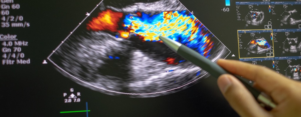
NDI doctors are U.S. Board Certified and hold certifications in echocardiography.
Remote Cardiology Services In The United States
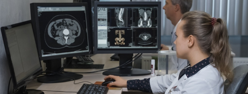
The National Diagnostic Imaging telecardiology company offers multiple remote diagnostic services online such as echocardiogram reading services and ECG interpretation services.
NDI telecardiology services are used by primary care physicians to electronically share cardiac imaging studies online with NDI cardiologists who write expert ECG interpretations and deliver actionable final reports that help to save lives.
The NDI telecardiology company provides remote online reporting of cardiac imaging studies of the circulatory system, cardiovascular system, heart and blood vessels.
NDI telecardiology services are a specialized version of telemedicine used by primary care physicians in the United States to help treat patients with cardiac problems.

Family physicians contract with telecardiology companies such as National Diagnostic Imaging to get access to telecardiology services in order to improve their treatment of patients with heart rhythm disorders (HRD) and cardiovascular disease.
NDI telecardiology services improve the speed, quality and cost-effectiveness of treating patients with cardiovascular disease on the primary level.
Cardiovascular disease (CVD) is a general term that describes a disease of the heart or blood vessels.
US Telecardiology Service To Diagnose Heart Disease
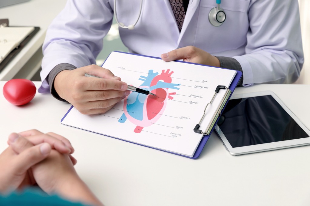
The NDI telecardiology company provides remote online reporting of cardiac imaging studies of the circulatory system, cardiovascular system, heart and blood vessels. NDI telecardiology services are used by U.S. cardiovascular imaging centers to receive remote interpretations of electrocardiogram (ECG or EKG) results from heart imaging specialists online.
Contact National Diagnostic Imaging For EKG Reading Services
Please complete the form below, call 216-514-1199 or email info@ndximaging.com to request a quote with the most competitive pricing in the industry.
If you are interested in a tele-echocardiography job, click here for information. Get more information on teleradiology pricing here.
To Request a Quote Complete The Form Below
How NDI Cardiologists Diagnose Heart Disease Online
Heart disease tests include echocardiograms, electrocardiograms, chest X-rays, CT scans, and MRI scans. An ECG is often used alongside other tests to help diagnose and monitor conditions affecting the heart. It can be used to investigate symptoms of a possible heart problem, such as chest pain, palpitations (suddenly noticeable heartbeats), dizziness and shortness of breath.
NDI cardiologists use a PACS workstation to interpret results from patient cardiac imaging studies, diagnostic radiology exams and diagnostic medical imaging tests.
PACS is used in cardiology departments. Cardiology has unique requirements; these needs come in the form of capturing sound, certain cardiac measurements, and structured cardiac catheter laboratory (cath-lab) and echocardiography laboratory reporting.
Cath-lab and echo are the chief modalities that are connected to a cardiology PACS. Others include cardiac magnetic resonance imaging (MRI) and cardiac computer tomography (CT). Cardiology PACS uses information produced by these 4 modalities.
Echocardiography, including stress echocardiography, can provide the required clinical information and avoids radiation exposure.
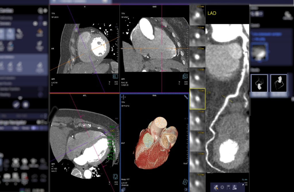
CT coronary angiography is increasingly used to detect coronary artery disease in patients with an intermediate risk and in those with equivocal stress test results.
Cardiac MRI studies are ordered by a patient’s cardiologist as an adjunct to other imaging modalities when further clarification is warranted. Cardiac MRI is an advanced imaging test used to obtain detailed views of the heart.
Once a cardiac image is generated, PACS is used to transfer that digital image to a computer network that will store the image and display the image for physicians. Learn more about PACS, computer networks and teleradiology in this PDF.
A picture archiving and communication system (PACS) is a high-speed, graphical, computer network system for the storage, recovery, and display of diagnostic radiological images.
NDI radiologists use PACS to diagnose heart disease, to dictate their findings and critical results, to write and sign their final radiology report and to deliver their conclusions in PDF format via teleradiology.
Cardiac imaging providers and treating physicians use high speed Internet connections to transmit pictures of a patient’s heart to the National Diagnostic Imaging PACS system.
Many different tests are used to diagnose heart disease. Besides blood tests and a chest X-ray, tests to diagnose heart disease can include:
- Electrocardiogram (ECG or EKG). An ECG is a quick and painless test that records the electrical signals in the heart. It can tell if the heart is beating too fast or too slowly.
- Holter monitoring. A Holter monitor is a portable ECG device that’s worn for a day or more to record the heart’s activity during daily activities. This test can detect irregular heartbeats that aren’t found during a regular ECG exam.
- Echocardiogram. This noninvasive exam uses sound waves to create detailed images of the heart in motion. It shows how blood moves through the heart and heart valves. An echocardiogram can help determine if a valve is narrowed or leaking.
- Exercise tests or stress tests. These tests often involve walking on a treadmill or riding a stationary bike while the heart is monitored. Exercise tests help reveal how the heart responds to physical activity and whether heart disease symptoms occur during exercise. If you can’t exercise, you might be given medications.
- Cardiac catheterization. This test can show blockages in the heart arteries. A long, thin flexible tube (catheter) is inserted in a blood vessel, usually in the groin or wrist, and guided to the heart. Dye flows through the catheter to arteries in the heart. The dye helps the arteries show up more clearly on X-ray images taken during the test.
- Heart (cardiac) CT scan. In a cardiac CT scan, you lie on a table inside a doughnut-shaped machine. An X-ray tube inside the machine rotates around your body and collects images of your heart and chest.
- Heart (cardiac) magnetic resonance imaging (MRI) scan. A cardiac MRI uses a magnetic field and computer-generated radio waves to create detailed images of the heart.
NDI Cardiologists Collaborate Online With Other Doctors
NDI cardiologists use teleradiology to receive radiological patient images 24/7 that are shared online by other radiologists and physicians.

NDI telecardiology services are used by U.S. cardiovascular imaging centers to receive remote interpretations of electrocardiogram (ECG or EKG) results from heart imaging specialists online.
Treating physicians use NDI telecardiology services to order and receive cardiac imaging study final reports written by NDI imaging cardiologists that are delivered in PDF format.
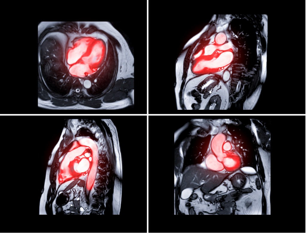
Cardiac magnetic resonance imaging (cardiac MRI or CMR) produces detailed images of the beating heart.
NDI cardiologists interpret cardiac magnetic resonance imaging (MRI) studies and state their cardiac MRI findings in reports that are provided online to healthcare providers and patients.
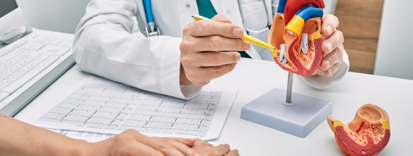
NDI diagnostic radiologists are licensed in all 50 states to use telecardiology services to collaborate with other physicians and to interpret cardiac imaging studies.
2025 NDI Telecardiology Reporting Services For Cardiac Imaging Studies
- Echocardiogram (echo).
- Single-photon emission computed tomography (SPECT).
- Cardiac positron emission tomography (PET).
- Cardiac computed tomography (CT).
- Nuclear cardiac stress test.
- Coronary angiogram or left heart catheterization.
- Cardiac MRI.
National Diagnostic Imaging provides telecardiology services, cardiac imaging interpretations, mobile EKG reporting services, remote cardiology reading services and echocardiogram (echocardiography) reading services in the USA.
Current Cardiovascular Imaging Reports
Current Cardiovascular Imaging Reports provides in-depth review articles contributed by international experts on the most significant developments in the field.
By presenting clear, insightful, balanced reviews that emphasize recently published papers of major importance, the journal elucidates current and emerging concepts regarding cardiovascular imaging techniques and technologies.
- Imaging in Cardiac Sarcoidosis: Complementary Role of Cardiac Magnetic Resonance and Cardiac Positron Emission Tomography
- The Clinical Role of 2D and Doppler Echocardiography Artifacts: a Review
- Echo-Based Hemodynamics to Help Guide Care in Cardiogenic Shock: a Review
- Update on CT Imaging of Left Ventricular Assist Devices and Associated Complications
- Insights into Myocardial Perfusion PET Imaging: the Coronary Flow Capacity
Circulation: Cardiovascular Imaging | AHA/ASA Journals
- December 21, 2022: Utilization of Cardiovascular Magnetic Resonance Imaging for Resumption of Athletic Activities Following COVID-19 Infection: An Expert Consensus Document on Behalf of the American Heart Association Council on Cardiovascular Radiology and Intervention Leadership and Endorsed by the Society for Cardiovascular Magnetic Resonance
- December 2, 2022: Imaging for Transcatheter Mitral Valve Edge-to-Edge Repair for an Unusual Cause of Cardiogenic Shock
- December 2, 2022: Fatal Pulmonary Embolism Resulting From a Popliteal Venous Aneurysm
- November 30, 2022: Isolated Left Ventricular Apical Hypoplasia: A Very Rare Congenital Anomaly Characterized by Multimodality Imaging and Invasive Testing
Accreditation Programs For Diagnostic Imaging Centers In The U.S.
Diagnostic imaging lets doctors look inside human and animal bodies for clues about a medical condition. A variety of machines and techniques can create pictures of the structures and activities inside the body. The type of imaging a doctor uses depends on the symptoms and the part of your body being examined. Ultrasonography is a popular diagnostic imaging tool that looks inside a dog or cat’s body via the use of sound waves.
ACR Accreditation is recognized as the gold standard in medical imaging. The ACR offers accreditation programs in CT, MRI, breast MRI, nuclear medicine and PET as mandated under the Medicare Improvements for Patients and Providers Act (MIPPA) as well as for modalities mandated under the Mammography Quality Standards Act (MQSA). Accreditation application and evaluation are typically completed within 90 days.
The ACR has accredited more than 39,000 facilities in 10 imaging modalities. They offer accreditation programs in Mammography, CT, MRI, Breast MRI, Nuclear Medicine and PET, Ultrasound, Breast Ultrasound and Stereotactic Breast Biopsy.
The Joint Review Committee on Education in Radiologic Technology (JRCERT) accredits educational programs in radiography, radiation therapy, magnetic resonance, and medical dosimetry.
The National Accreditation Program for Breast Centers (NAPBC) provides the structure and resources you need to develop and operate a high-quality breast center. Programs that are accredited by the NAPBC follow a model for organizing and managing a breast center to facilitate multidisciplinary, integrated, comprehensive breast cancer services.
Get information from the Centers for Medicare & Medicaid Services (CMS) about their requirements for accreditation of advanced diagnostic imaging suppliers, here.
The Intersocietal Accreditation Commission (IAC) is a nonprofit, nationally recognized accrediting organization. The IAC was founded by medical professionals to advance appropriate utilization, standardization and quality of diagnostic imaging and intervention-based procedures.
The IAC is a nonprofit organization in operation to evaluate and accredit facilities that provide diagnostic imaging and procedure-based modalities, thus improving the quality of patient care provided in private offices, clinics and hospitals where such services are performed.
With a 30-year history of offering medical accreditation to facilities within the U.S. and Canada, IAC is also now offering accreditation in international markets. The IAC programs for accreditation are dedicated to ensuring quality patient care and promoting health care and all support one common mission: Improving health care through accreditation®.
The ACVR is the American Veterinary Medical Association (AVMA) recognized veterinary specialty organization™ for certification of Radiology, Radiation Oncology and Equine Diagnostic Imaging.
If you are a radiology imaging service in the United States that is looking for a company that can provide teleradiology coverage for your current and future case volume, contact National Diagnostic Imaging by phone at 216-514-1199 or by emailing info@ndximaging.com.
Imaging Facilities Accredited by the American College of Radiology
Use this search form to find imaging facilities accredited by the American College of Radiology.
Facilities: To verify the accreditation status of specific units within your imaging facility, please call 1-800-770-0145.
ACR Accredited Facility Designations
Video Posted On You YouTube.com On November 11, 2015 By RadiologyACR
