NIOSH-Certified B Read Service Rates From $40 Per Study
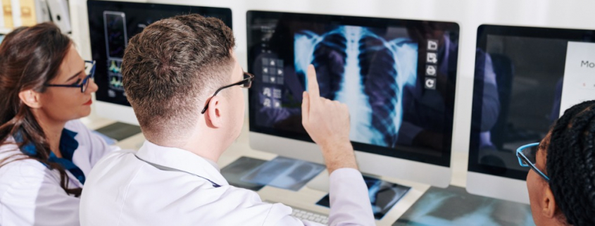
In 2025, NDI B-readers provide sophisticated and detailed interpretations of chest x-rays and CT scans, as well as screening chest radiographs for pneumoconiosis on behalf of occupational health clinics with B read programs that provide expert respiratory disease diagnosis and treatment for black lung disease.
B Read Services For Occupational Health Clinics And Programs In The US
Occupational clinics, doctors and attorneys work with experienced NIOSH certified B readers at the National Diagnostic Imaging company for the detection and surveillance of occupational lung disease.
Choosing a highly qualified lung imaging radiologist to detect black lung disease can make a difference. Specialized lung imaging radiologists at NDI know the anatomy of the lungs exceptional well.
Medical Imaging For Black Lung Disease
In 1969, the U.S. Congress passed a new piece of legislation on coal mining that called for compensation to miners who had developed pneumoconiosis, or black lung.
The Black Lung Benefits Act was originally enacted by Congress in 1969 as part of the Federal Coal Mine Health and Safety Act to provide benefits to coal miners disabled by pneumoconiosis, or black lung disease.
The new program relied heavily on medical imaging to detect black lung disease, with radiology playing a major role in the program’s implementation.
The legislation directed the U.S. Public Health Service to establish a system to detect respiratory difficulties as shown on chest x-ray for individual miners.
Fees For B Reads for Chest X-Rays For Occupational Health Start At $40 Per Study
Chest X-rays are the primary method for diagnosing black lung disease, also known as coal workers’ pneumoconiosis (CWP). A chest X-ray can show the presence of scar tissue or inflammation in the lungs. A CT scan can also reveal scar tissue in the lungs.
For more information about occupational and black lung disease, watch this video Dr Catherine Jones – Black Lung and the Radiology Revolution.
Specially qualified NDI radiologists examine x-rays of the lungs to look for signs of pneumoconiosis, also known as black lung disease.
Chest radiography is a standard tool used in medical screening for pneumoconiosis. The chest radiograph remains the primary mode of screening for pneumoconiosis in the United States and elsewhere.
NDI B readers are radiologists that are certified by the National Institute of Occupational Health and Safety (NIOSH).
NIOSH Certified B Readers And NIOSH B Reader List
B Readers at NDI classify chest radiographs of workers exposed to mineral dusts. They use a specific protocol to read and record their findings, which is developed by the International Labour Organization (ILO).
Certification through NIOSH’s B Reader program indicates proficiency in classifying posteroanterior chest radiographs using the ILO system.
The ILO International Classification of Radiographs of Pneumoconioses provides a means for describing and recording systematically the radiographic abnormalities in the chest provoked by the inhalation of dusts. Learn more about imaging to diagnose occupational lung disease, by watching this radiology YouTube video.
A list of approved providers for the federal Black Lung Program is available from the U.S. Department of Labor in PDF format, here.
Physicians from inside the United States that have who have demonstrated competence in applying the ILO classification by successfully completing the NIOSH B Reader examination.
The NIOSH B Reader list is available online, here.
The Importance Of B Read Services Offered By NDI
- B reads provided by NDI radiologists assist professionals in the occupational health industry to provide medical surveillance for workers exposed to respirable mineral dusts, such as asbestos, coal mine dust, and crystalline silica.
- NDI’s B reads help government agencies track the population burden of pneumoconiosis.
- NDI provides B reading services for professionals that participate in legal and administrative activities, such as compensation programs.
In 2025, National Diagnostic Imaging provides b-reading reports and interpretations online via teleradiology services. NDI provides B read x-ray interpretations for physicians, coal workers and asbestos medical surveillance programs.
Other NDI B read clients include insurance companies, attorneys, industry-sponsored medical screening organizations and medical organizations that are concerned with occupational respiratory diseases.
To find an NIOSH approved x-ray facility that is close to you, use this map.
Request NIOSH-Certified B Reader Services From NDI
Call 216-514-1199 To Request A B Reading Service Quote Or Complete The Form Below
If You Want Access To A NIOSH Certified B Reader Or You Want A Quote For B Reads, Please Complete The Form Below

Expert “B” Readers from the National Diagnostic Imaging company are certified by the National Institute for Occupational Safety and Health (NIOSH). B read reports from specialized NDI lung imaging radiologists can document whether a patient may be eligible for federal programs and benefits due to black lung disease. B-reader fees start at $40 per report.
Accurate And Timely B-Reader Services Are Performed By NDI Radiologists Certified By NIOSH
NDI B readers provide comprehensive and detailed interpretations of chest X-rays and chest CT scans for occupational health and litigation screenings.
Chest radiographs must be interpreted and classified in accordance with the Guidelines for the Use of the ILO International Classification of Radiographs of Pneumoconioses.
A chest radiograph (X-ray) must be of suitable quality for proper classification of pneumoconiosis and must conform to the standards for administration and interpretation of chest X-rays as described in Appendix A, here.
Lung imaging plays an essential role in evaluating lung diseases such as black lung disease.
Review the NIOSH B Reader list, here. Learn about certified B Reader radiologists here.
Learn about NIOSH B reader training and examinations, here. Review the NIOSH B Reader syllabus, here.
In 2025, the easiest way to get a B read or b-reading interpretations in the United States is to call NDI at 1-800-950-5257 or email info@ndximaging.com.
NDI B Readers Are Competent In Radiographic Reading And Proficient In The Classification Of X-Rays Of Pneumoconiosis
NIOSH B readers at the National Diagnostic Imaging teleradiology service, examine x-rays of the lungs to find indications of pneumoconiosis, a lung disease known as “Black Lung Disease”.
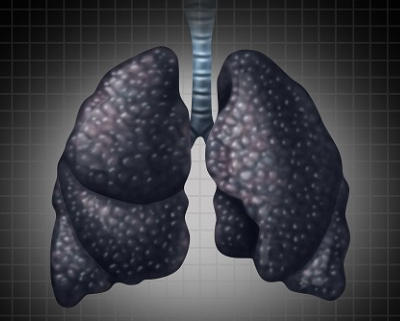
Coal workers’ pneumoconiosis (CWP), commonly known as “black lung disease,” occurs when coal dust is inhaled. Over time, continued exposure to the coal dust causes scarring in the lungs, impairing your ability to breathe. Considered an occupational lung disease, it is most common among coal miners.
A B reading on a chest x-ray looks for changes on the chest x-ray that may prove exposure and disease caused by particles such as asbestos and silica.
A B Reader is a physician certified by the National Institute for Occupational Safety and Health (NIOSH) as demonstrating proficiency in classifying radiographs of the pneumoconiosis.
Pneumoconiosis is a lung disease caused by breathing in certain kinds of dust particles that damage your lungs.
The B Reader examination was initially developed to identify physicians qualified to serve in national pneumoconiosis programs directed at coal miners and others who suffer from dust-related illness.
The B Reader Program aims to ensure competency in radiographic reading by evaluating the ability of readers to classify a test set of radiographs.
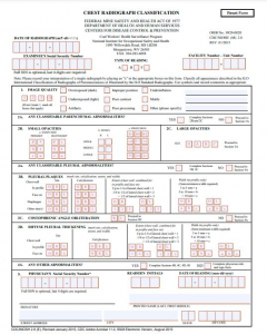
Download a NIOSH B Reader form (Radiograph Interpretation Form) used by the Coal Workers’ Health Surveillance Program in PDF format, here.
B Read X-rays Are Sophisticated Interpretations Of Chest X-Rays
The qualified “B” readers at NDI are radiologists that are certified by the National Institute for Occupational Safety and Health (NIOSH) as demonstrating proficiency in classifying radiographs of the pneumoconiosis.
Physicians at NDI are qualified to serve in national pneumoconiosis programs directed at coal miners and others who suffer from dust-related illness. NDI physicians who read chest X-rays for work-related diseases like black lung are known as “B readers.”
Chest Radiography And Pneumoconiosis Chest X-Rays
Chest radiography is a principal tool in the diagnosis of pneumoconiosis.
Pneumoconioses are a broad group of lung diseases that result from inhalation of dust particles.
An x-ray may be helpful in the diagnosis of pneumoconiosis. Findings on an x-ray suggestive of/diagnostic of pneumoconiosis include:
- Pleural plaques are pathognomonic for asbestosis.
- Interstitial nodules in the upper zone of the lungs for both silicosis and coal worker’s pneumoconiosis.
- Progressive massive fibrosis is seen in both silicosis and coal worker’s pneumoconiosis.
- Hilar adenopathy with ground glass opacities is seen in berylliosis.
Pneumoconiosis, Causes, Signs and Symptoms, Diagnosis and Treatment.
Video Posted To YouTube on February 24, 2021 by Medical Centric
Clinical picture: Digging into coal workers pneumoconiosis
Video Posted On YouTube.com on July 3, 2020 by Lancet
NDI B readers are certified by the National Institute for Occupational Safety and Health (NIOSH) for both federal and state compensation claims. B readers do not specifically have to be pulmonologists or radiologists, though they can be both.
US licensed physicians who achieve a score of 50% or more on the initial examination are certified as NIOSH B Readers. B Readers are certified for 4 years from the date of approval, after which passing the recertification examination is required to maintain the B Reader certification.
The easiest way to get access to teleradiology services for B reading services in the United States is to call National Diagnostic Imaging at 1-800-950-5257 or email info@ndximaging.com.
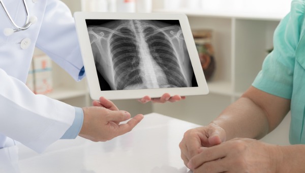
The B-reading is a special reading of a standard chest x-ray film performed by a physician certified by the National Institute for Occupational Safety and Health (NIOSH). The reading looks for changes on the chest x-ray that may indicate exposure and disease caused by agents such as asbestos or silica.
Certified B reader consultants, certified B reader radiologists, NIOSH-certified B readers and lung imaging radiologists work at National Diagnostic Imaging.
B Read x-rays are a sophisticated interpretation of a chest x-ray. NDI B readers identify exposure and illness caused by substances such as asbestos and silica.
Strict guidelines for B read x-rays provide protocols for examining x-rays and recording changes/anomalies on chest x-rays that may be caused by fibers and dust inhalation.
For information on the B Reader Program and Certification Course, NIOSH B Reader Training and Examination, click here.
For new development impacting the B Reader Program, click here. For information on the ACR Lung Cancer Screening Registry, click here.
For information on ethical considerations for B Readers, click here. Physicians interested in becoming NIOSH certified B readers or current B readers interested in updating their practice can get more information here.
Classifying And Detecting Occupational Lung Diseases
Chest radiographic imaging (chest x-ray) is a widely applied and important tool for assessing lung health in workers exposed to dusts capable of producing pneumoconiosis and other diseases. Chest radiographs are the most common film taken in medicine.
Pneumoconiosis includes asbestosis, silicosis and coal workers’ pneumoconiosis (CWP). CWP is sometimes called “Black Lung Disease“. Silicosis is a disease caused by inhaling respirable silica dust. Asbestosis is a chronic lung disease caused by inhaling asbestos fibers.
NDI offers consulting services and reviews of chest radiographs or CT scans for the presence of occupational lung disease.
National Diagnostic Imaging utilizes chest radiographs, teleradiology, digitally-acquired chest radiographs, x-rays, CT scans and digital imaging to classify, recognize and detect signs of occupational lung disease, interstitial lung disease, pneumoconiosis/occupational disease, illnesses relating to asbestos silica exposure, silicosis and asbestosis.
About the Black Lung Program
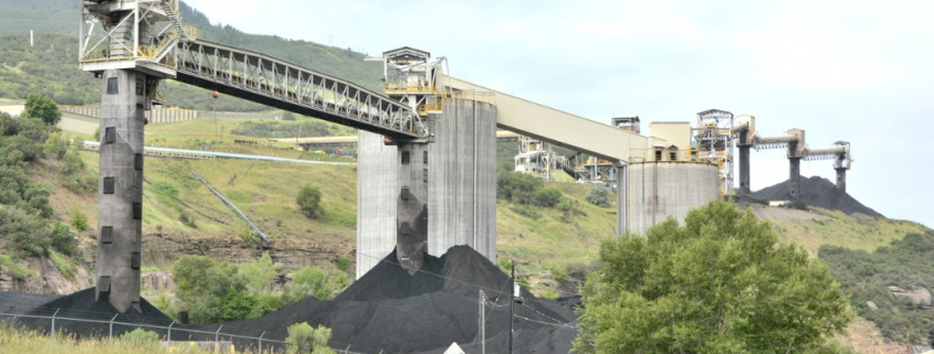
The Division of Coal Mine Workers’ Compensation, or Federal Black Lung Program, administers claims filed under the Black Lung Benefits Act. The DCMWC provides monthly payments and medical benefits to eligible coal miners who are totally disabled due to black lung disease.
The Act provides compensation to coal miners who are totally disabled by pneumoconiosis arising out of coal mine employment, and to survivors of coal miners whose deaths are attributable to the disease. The Act also provides eligible miners with medical coverage for the treatment of lung diseases related to pneumoconiosis.
The Black Lung Benefits Act provides monthly payments and medical benefits to coal miners totally disabled from pneumoconiosis (black lung disease) arising from their employment in or around the nation’s coal mines. The Act also provides monthly benefits to a miner’s dependent survivors.
About Pulmonary Fibrosis
Pulmonary fibrosis is a lung disease that occurs when lung tissue becomes damaged and scarred. This thickened, stiff tissue makes it more difficult for your lungs to work properly. As pulmonary fibrosis worsens, you become progressively more short of breath.
Pulmonary fibrosis (PF) may be difficult to diagnose as the symptoms of PF are similar to other lung diseases. There are many different types of PF.
Tests like chest X-rays and CT scans can help doctors look at lungs to see if there is any scarring.
- Many people with PF actually have normal chest X-rays in the early stages of the disease.
- A high-resolution computed tomography scan, or HRCT scan, is an X-ray that provides sharper and more detailed pictures than a standard chest X-ray and is an important component of diagnosing PF.
- Your doctor may also perform an echocardiogram (ECHO).
- This test uses sound waves to look at your heart function.
- Doctors use this test to detect pulmonary hypertension, a condition that can accompany PF, or abnormal heart function.
Pneumoconiosis: Pulmonary Fibrosis Caused by Occupational Exposures
Posted On YouTube On March 5, 2020 by Pulmonary Fibrosis Foundation
Expert Witnesses, Expert Medical Testimony And Litigation Support For Asbestosis, Silicosis And Other Occupational Dust And Fiber Exposures
Radiologists at NDI are Federal Government Certified B-Readers for asbestosis, silicosis and other occupational dust and fiber exposures.
The radiologists at NDI assist lawyers and insurance companies with their B-reading related litigation needs including reading any CT scans for pneumoconiosis.
They are experienced as expert witnesses and medicolegal consultants (both defense and plaintiff) and are prompt, reliable, and efficient, having consulted in dozens of cases.
Request B Reader Services, Fees And Rates
If you require the services of a NIOSH-certified B reader please complete the form below, call 216-514-1199 or email info@ndximaging.com to request a quote.
Rates are typically well below other service providers. We competitively price these studies per read and total pricing is based on the number of reads requested and other details involved in providing B Reader Services.
Accreditation Programs For Diagnostic Imaging Centers In The U.S.
Diagnostic imaging lets doctors look inside human and animal bodies for clues about a medical condition. A variety of machines and techniques can create pictures of the structures and activities inside the body. The type of imaging a doctor uses depends on the symptoms and the part of your body being examined. Ultrasonography is a popular diagnostic imaging tool that looks inside a dog or cat’s body via the use of sound waves.
ACR Accreditation is recognized as the gold standard in medical imaging. The ACR offers accreditation programs in CT, MRI, breast MRI, nuclear medicine and PET as mandated under the Medicare Improvements for Patients and Providers Act (MIPPA) as well as for modalities mandated under the Mammography Quality Standards Act (MQSA).
Accreditation application and evaluation are typically completed within 90 days.
The ACR has accredited more than 39,000 facilities in 10 imaging modalities. They offer accreditation programs in Mammography, CT, MRI, Breast MRI, Nuclear Medicine and PET, Ultrasound, Breast Ultrasound and Stereotactic Breast Biopsy.
The Joint Review Committee on Education in Radiologic Technology (JRCERT) accredits educational programs in radiography, radiation therapy, magnetic resonance, and medical dosimetry.
The National Accreditation Program for Breast Centers (NAPBC) provides the structure and resources you need to develop and operate a high-quality breast center.
Programs that are accredited by the NAPBC follow a model for organizing and managing a breast center to facilitate multidisciplinary, integrated, comprehensive breast cancer services.
Get information from the Centers for Medicare & Medicaid Services (CMS) about their requirements for accreditation of advanced diagnostic imaging suppliers, here.
The Intersocietal Accreditation Commission (IAC) is a nonprofit, nationally recognized accrediting organization.
The IAC was founded by medical professionals to advance appropriate utilization, standardization and quality of diagnostic imaging and intervention-based procedures.
The IAC is a nonprofit organization in operation to evaluate and accredit facilities that provide diagnostic imaging and procedure-based modalities, thus improving the quality of patient care provided in private offices, clinics and hospitals where such services are performed.
With a 30-year history of offering medical accreditation to facilities within the U.S. and Canada, IAC is also now offering accreditation in international markets.
The IAC programs for accreditation are dedicated to ensuring quality patient care and promoting health care and all support one common mission: Improving health care through accreditation®.
The ACVR is the American Veterinary Medical Association (AVMA) recognized veterinary specialty organization™ for certification of Radiology, Radiation Oncology and Equine Diagnostic Imaging.
If you are a radiology imaging service in the United States that is looking for a company that can provide teleradiology coverage for your current and future case volume, contact National Diagnostic Imaging by phone at 216-514-1199 or by emailing info@ndximaging.com.
Imaging Facilities Accredited by the American College of Radiology
Use this search form to find imaging facilities accredited by the American College of Radiology.
Facilities: To verify the accreditation status of specific units within your imaging facility, please call 1-800-770-0145.
ACR Accredited Facility Designations
Video Posted On You YouTube.com On November 11, 2015 By RadiologyACR

